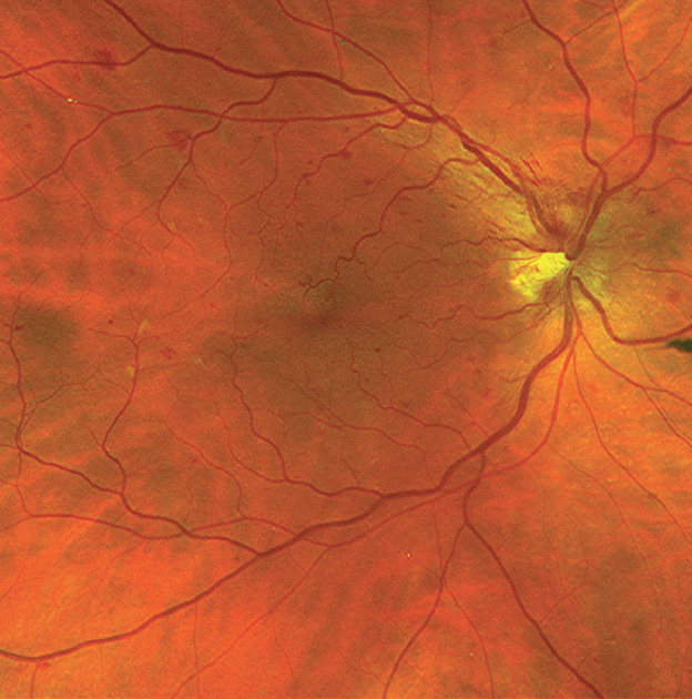
1-7 At first, it was believed to be solely of hypertensive origin however, this was later refuted when diabetic patients without hypertension also presented with a CWS.įurthermore, a CWS may be a retinal characteristic in patients with AIDS, anemia, collagen disease, bacterial endocarditis, radiation retinopathy, vascular occlusive disease, leukemia and systemic lupus erythematosus. In most cases, the finding is related to systemic disease. Microaneurysms, dot and blot hemorrhages, intraretinal microvascular abnormalities, neovascularization of the disc or retina, venous beadingĪ CWS may present as a solitary retinal finding or be accompanied by additional pathology that can contribute to the overall clinical picture. 4ĭISTINGUISHING FEATURES OF THE TWO MOST COMMON CAUSES OF COTTON WOOL SPOTSĪrteriolar narrowing, arteriovenous nicking, flame hemorrhages, microaneurysms, disc edema Second, the disruption of flow is presumed to be from an obstructed arteriole, in some cases, thus impairing normal vessel filling. First, the presence of the CWS, to some extent, masks the underlying fluorescence. The reason for the darkened area is twofold.

In contrast to OCT, fluorescein angiography (FA) of a CWS manifests as a focal area of hypofluorescence that corresponds with the lesion. Following clinical resolution of the CWS, retinal thinning may be evident in the corresponding area. A scan over the area of the CWS will reveal marked thickening and hyper-reflectivity of the RNFL and inner neurosensory retina. Since these lesions accumulate within the RNFL, the abnormality on OCT is typically found within this inner retinal layer. 3īesides the typical funduscopic appearance, a CWS can also be identified and monitored using optical coherence tomography (OCT). 1 Although patients are typically asymptomatic, some reports indicate either local or arcuate scotomas at the site of the CWS. 4 Because the RNFL thickens as we move toward the periphery of the retina, a cotton wool spot is most evident within the post-equatorial region. The “cytoid body,” or end bulb of Cajal, is the histological landmark of the CWS and represents the terminal swelling of an RGC axon from the disruption of flow and hypoperfusion of the underlying retina. 1,3ĭiabetic retinopathy and a CWS along the superior temporal RNFL arcade. The CWS is a manifestation of this interruption of flow and is an accumulation of axoplasmic debris within the unmyelinated axons.

In the ischemic retina, both orthograde and retrograde axonal transport of RGCs is disrupted. The retina is comprised of several kinds of neurons, including the retinal ganglion cells (RGCs), which are located within the superficial retinal nerve fiber layer (RNFL). Deep DiveĪ CWS is thought to originate in cases where there is reduced perfusion, leading to focal or diffuse retinal ischemia. To understand the health implications that cotton wool spots may carry, it is necessary to explore the process behind their formation. 1,2 This small but important finding can be a marker for potentially life-threatening conditions, making it of great clinical utility.

A CWS appears as a white and fluffy superficial lesion 0.1mm to 1.0mm in diameter that obscures the underlying retinal detail. One of these potential retinal findings is the cotton wool spot (CWS). A careful retinal examination is critical in order to evaluate each patient for various pathologies, some of which yield an underlying systemic cause.


 0 kommentar(er)
